But identifying dead cells can be tricky and has been a constant problem throughoutmy career as a neuroscientist.
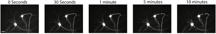
Dead cells have acharacteristic balled-up appearancethat is relatively easy to recognize once you know what to look for.
Unfortunately, doing this by hand is a slow, expensive, and sometimes error-prone process.

This is a time-lapse of what a dying neuron looks like under a microscope.
It became clear to us that manual curation was neither accurate nor efficient.
My colleagues and I have been trying for some time to automate the curation process.
But anew artificial intelligence technologymy research team developed can identify dead cells with both superhuman accuracy and speed.
This advance could potentially turbocharge all kinds of biomedical research, especially on neurodegenerative disease.
AI to the rescue
Artifical intelligencehas recently taken the field of microscopy by storm.

It’s free, every week, in your inbox.
Convolutional neural networks can be trained to recognize and discover complex patterns in images.
These patterns could includebiological phenomenadifficult to see by eye.
For example, one research group was able to train CNNs to identifyskin cancermore accurately than trained dermatologists.
First, we needed to teach the BO-CNN to distinguish between clearly dead and clearly alive cells.
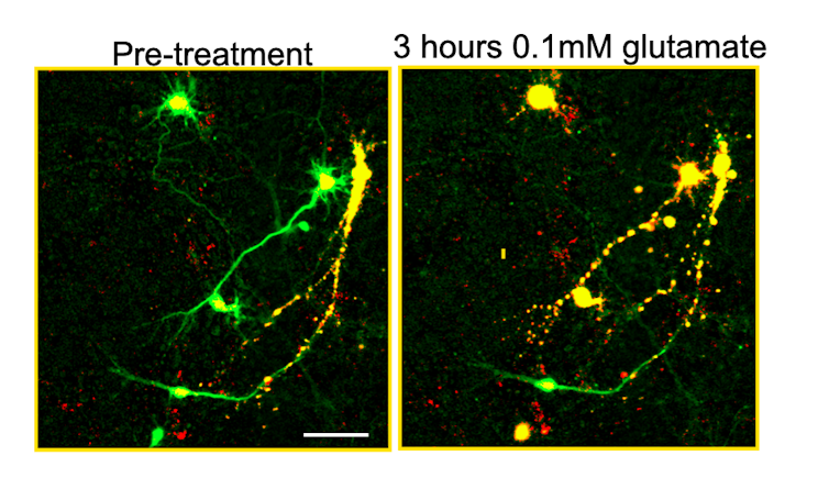
The BO-CNN could easily learn that green meant alive and yellow meant dead.
One obvious question still remained, however why was our model so effective at finding dead cells?
To figure out what these patterns were, we used additionalcomputational toolsto create visual representations of the BO-CNNs decisions.
These images show alive neurons colored green and dead neurons colored yellow.
Live-cell microscopy is extremely rich with information that researchers have difficulty interpreting.
